2 The Scientific Foundation: What the Evidence Reveals
In Chapter 1, we explored how the retina serves as a remarkable window into human health. We discussed its unique anatomical and physiological properties, its connection to the brain, and the various eye pathologies visible through traditional fundus photography. This chapter takes the next step—examining how artificial intelligence has transformed retinal imaging from a specialized diagnostic tool into a powerful platform for comprehensive health assessment.
For decades, retinal assessment was limited by human perceptual abilities. Even highly trained ophthalmologists were constrained by what the human eye could discern and what the human brain could process. Subtle vascular changes, minute tissue alterations, and complex pattern relationships often remained invisible or unrecognized. The introduction of artificial intelligence has fundamentally changed this paradigm.
Where traditional diagnostics relied on identifying known pathological features—the enlarged optic cup of glaucoma or the distinctive exudates of diabetic retinopathy—AI systems can detect statistical patterns and relationships that have no established visual correlates. These systems don’t just see differently; they see more, analyzing thousands of parameters simultaneously and identifying correlations invisible to human observers.
This transformation mirrors the broader evolution in medical diagnostics—from reactive identification of established disease to proactive recognition of health trajectories and risk factors. The evidence we’ll explore in this chapter demonstrates how AI-powered retinal analysis moves beyond traditional diagnostics toward a new model of health assessment: more accessible, more comprehensive, and more predictive than previously possible.
2.1 Artificial Intelligence: The Breakthrough Enabler
The true power of retinal imaging emerges through the application of advanced artificial intelligence. While human experts can identify obvious retinal pathologies, AI systems can detect subtle patterns and correlations invisible to the human eye.
Deep Learning Architecture
Modern retinal analysis systems employ deep learning networks—sophisticated AI architectures inspired by the human brain’s neural structure. These networks contain multiple processing layers that progressively extract higher-level features from raw image data.
During the training process, these networks learn to identify patterns by analyzing millions of retinal images paired with known health outcomes. The system gradually develops the ability to recognize subtle relationships between retinal features and various health conditions. This learning process goes far beyond simple pattern matching—it enables the AI to discover new biomarkers and relationships that might never have been identified through traditional research methods.
What distinguishes deep learning from previous computational approaches is its ability to automatically discover relevant features without explicit programming. Traditional image analysis might require engineers to specify exactly what features to measure (like vessel width or branching patterns). In contrast, deep learning systems determine independently which image characteristics are most relevant for health assessment, often identifying patterns too subtle or complex for human observers to recognize.
Quantitative Analysis Capabilities
AI-powered retinal analysis can precisely quantify numerous parameters that would be challenging for human assessment:
Vascular Measurements: Automatic measurement of arteriolar and venular caliber, tortuosity, branching angles, and vessel wall characteristics with micrometer precision.
Structural Quantification: Analysis of optic disc parameters (cup-to-disc ratio, neuroretinal rim area), macular region characteristics, and nerve fiber layer integrity.
Textural Analysis: Evaluation of retinal background texture and subtle changes in reflectance that may indicate early pathological changes.
Longitudinal Comparisons: Precise tracking of changes over time, allowing for early detection of progressive conditions and monitoring of treatment responses.
Comparative Analysis: Matching patient findings against large normative databases, considering factors like age, sex, and ethnicity to provide contextualized health insights.
These quantitative capabilities transform retinal imaging from a simple screening tool into a sophisticated health assessment platform capable of detecting subtle variations that correlate with systemic health conditions.
2.2 The Advantage of Fundus Photography
While these concepts may be complex, they are made accessible through the use of high-resolution fundus photography. This specialized imaging technique uses a camera with particular optics and light spectrum to capture detailed images of the retina, including the optic disc, blood vessels, and the macula, the area of sharp central vision. These photographs reveal subtle changes not easily visible during a traditional ophthalmoscopy exam (looking into the eyes using a handheld tool). These photos also generate a permanent record of the retina that can be analysed by human and AI.
Through the lens of high-resolution fundus photography, AI can detect a host of changes in the retina that indicate the presence of an underlying condition. These changes may be related to:
- Vessel Caliber: The diameter of blood vessels (arterioles and venules). Narrowing or widening can be indicators of high blood pressure or inflammation.
- Vessel Tortuosity: The degree of bending or twisting in blood vessels. Abnormally tortuous vessels can be linked to age or other disease processes.
- Changes in Colour: Differences in the appearance of the retina related to the blood flow, oxygenation, or the presence of certain pigments.
- Presence of Lesions: Such as hemorrhages, exudates, and drusen.
- Changes in Retinal Layer Thickness: Variations in layer thickness have been shown to correlate with a variety of systemic diseases.
The power of fundus photography as a tool lies in its ability to capture these subtle signals, offering a glimpse into the intricate workings of the body. It reveals information not readily apparent through routine physical exams or blood tests. Often, these subtle retinal changes precede more pronounced systemic symptoms and can therefore act as a warning system, allowing for earlier detection and intervention.
This also highlights the importance of looking beyond the overt signs of disease. Many individuals, especially those interested in wellness and preventive medicine, may not have obvious symptoms of any disease and are considered “healthy”. However, even if a disease is yet to manifest clinically, subclinical or pre-symptomatic stages may be detectable via these subtle changes in retinal morphology.
By studying the retina through high-resolution fundus photography, we are no longer confined to assessing just eye-related health. Instead, this technology allows us to unlock the secrets held within this unique tissue. It enables us to:
- Assess the health of your vascular system
- Determine your likelihood of certain systemic conditions
- Gain insight into your biological age
- Identify potential early signs of disease
This holistic view, enabled by retinal imaging, moves us away from a purely reactive approach to health and towards a more proactive and personalized model of care. It is a method that acknowledges the interconnectedness of the body’s systems and empowers individuals to take control of their own health and wellbeing.
The retina, once considered solely as an organ of vision, is now recognized as a fascinating and valuable tissue that can be used to assess overall health. High-resolution fundus photography and the power of AI have given us the ability to delve into these secrets to find clues about disease states, and biological aging, opening up a new frontier in the way we understand, monitor and promote health. While research is ongoing, the basic principles and studies outlined in this section clearly demonstrate the amazing potential of this modality to revolutionize wellness and health assessment.
2.3 Deep Learning & Artificial Intelligence
The human eye is remarkable in its ability to discern incredibly subtle patterns. However, even the most skilled human eye can’t compete with the power of a computer when it comes to quickly processing and analyzing vast amounts of complex information. This is where deep learning and artificial intelligence (AI) become invaluable tools in the realm of high-resolution fundus photography. To fully appreciate the power of fundus imaging for health assessment, understanding the role of AI is essential.
As we discussed in the previous chapter, the retina holds a wealth of information about our overall health. However, identifying and interpreting the subtle changes within a retinal image can be challenging. This is where traditional methods have their limitations; relying on human interpretation is not only time-consuming, but it can also be subject to inter- and intra-reader variability (that is, one person might interpret the same photo differently on different occasions, and two people might interpret the same photo differently from each other). With AI, specifically deep learning, these limitations can be overcome.
Traditional computer programs have often relied on “hand-crafted” algorithms. These were built by human engineers, who would pre-program all the steps the program must take, and which features it should look for in the images. Deep learning provides a radical shift, because it is not programmed to follow pre-determined instructions. Instead, a deep learning system is trained on vast amounts of data. For example, instead of telling the AI program how to identify a blood vessel, a deep learning system is trained on hundreds of thousands of retinal images and their corresponding health outcomes, learning the subtle and complex relationships between image patterns and disease states. This process allows AI to detect patterns and features that a human eye might miss, making the diagnostic and predictive capabilities of fundus imaging even more powerful.
Deep learning is a specific type of machine learning (a subfield of AI) that employs artificial neural networks with multiple layers (hence the name “deep”). These layers enable the AI system to process information through hierarchical stages, similar to the complex networks in the brain. In effect, the AI algorithm “learns” what features are relevant for the task at hand, and “decides” on the relative weighting that should be applied to these features. In general, a deep learning model is trained on hundreds of thousands (or even millions) of retinal images with their corresponding ground-truth clinical diagnoses and other health information; thus, each layer in the neural network learns increasingly more abstract and relevant features, ultimately allowing it to perform a task as sophisticated as detecting glaucoma or diabetic retinopathy, or predicting an individual’s biological age.
The advantage of this “deep” architecture is that it enables the AI model to automatically extract higher level and more nuanced characteristics from the images. For example, rather than being programmed to analyze just vessel caliber or vessel tortuosity, AI will automatically learn to assess these factors, and other image features, and then learns how to weigh these factors relative to the health outcomes. This also means that deep learning is capable of extracting new information, even about those underlying factors that may not even be discernable to the human eye, and which might have been missed using standard methods.
Opticare has incorporated this revolutionary technology into an AI-powered fundus camera to provide state-of-the-art health assessments. The Opticare AI system is a deep learning model trained on a massive dataset of millions of labeled retinal images. This training enables the system to identify subtle patterns in your retinal images and compare these patterns to known characteristics of different health states.
When you take a fundus photo with your Opticare device, the image is immediately analysed by a trained AI system. This system isn’t just looking at the obvious features of the retina; it is trained to assess everything the human eye can see, as well as the things the human eye can’t see. Some of these characteristics are as follows:
- Vessel Caliber and Tortuosity: AI accurately measures the diameter and shape of the blood vessels, which can be indicators of cardiovascular risk, diabetes, and other systemic conditions.
- Layer Thickness: The deep learning model is capable of analysing the thickness of different retinal layers, and any changes of those thicknesses over time or compared to a healthy population. Subtle differences in layers are often correlated with various diseases or risks of developing them.
- Presence of Lesions: AI can automatically identify various abnormal lesions such as drusen, hemorrhages and exudates, often signs of eye disease and also correlated with systemic disease.
- Color Changes: AI can detect subtle changes in the colour of the retina, which may indicate underlying conditions related to blood flow and metabolism.
- Spatial Organization of Features: Deep learning networks can discern patterns of how different features are spatially organized and how those patterns might be related to specific conditions, going beyond the assessment of single features alone.
- Combinations of Features: The AI models are trained to evaluate combinations of features, just like clinicians do, in order to arrive at a final diagnostic or risk evaluation. This approach takes advantage of the redundancy of the retinal features, and is more robust than relying on single features alone.
When using the Opticare camera, you should keep the following in mind:
Image Capture: A high-resolution fundus photograph of the retina is captured using a specialized camera and lighting.
Automated Analysis: This image is fed into our deep learning system which analyzes over 30 million retinal images during training.
Interpretation and Insights: The system provides an assessment of the retina as a marker for various diseases, and also generates a prediction of the biological age. You will be able to see a score or a graph that displays the findings clearly and simply.
Clinical Context: The AI’s findings should be used as an adjunct to your current clinical assessment and within the context of the specific client, rather than being a standalone diagnostic tool. A human expert should also interpret the findings, to ensure the best level of care.
Deep learning and AI are transforming the way we analyze fundus images. This technology empowers you to quickly identify subtle patterns that are not readily visible to the human eye. The Opticare AI fundus camera harnesses this power and provides a cutting-edge means for you to offer comprehensive and state-of-the-art health assessments to your clients. By bridging the gap between the complexity of retinal data and readily interpretable results, Opticare brings a new level of clarity, insight and value to your wellness practice.
2.4 A Powerful Tool for Early Diabetes Detection
Diabetes mellitus, particularly type 2 diabetes, represents one of the most significant global health challenges of our time, affecting hundreds of millions worldwide. Its impact extends far beyond glucose metabolism, affecting nearly every organ system in the body, including the eyes. The retina, with its unique and readily observable microvasculature, provides an extraordinary non-invasive window into both the presence and potential development of diabetes. Modern high-resolution fundus photography, combined with artificial intelligence (AI), is revolutionizing diabetes detection, enabling earlier identification and potentially improving patient outcomes.
The diabetic retina tells a story long before other symptoms emerge. Before the onset of full-blown diabetes, subtle changes occur in the retina, typically related to the impact of hyperglycemia on the microvasculature. These early changes often precede noticeable symptoms, making them invaluable for early detection. The earliest signs include vessel narrowing, where high blood sugar damages the delicate walls of retinal arterioles, reducing their diameter and blood flow. As vessel walls become compromised, they may leak fluid and blood components into the retinal tissue, leading to subtle swelling or the appearance of small, dot-like hemorrhages and exudates. Microaneurysms, appearing as tiny red dots, indicate damage to blood vessel walls, while changes in vessel color reflect alterations in blood flow and oxygenation. Additionally, research has shown that subtle changes in retinal layer thickness, particularly in the ganglion cell layer and inner plexiform layer, correlate with early diabetic retinopathy.
Traditional diabetes screening methods often prove invasive, time-consuming, and costly. Patients typically undergo fasting blood glucose tests, hemoglobin A1c measurements, or oral glucose tolerance tests, requiring specialized equipment, personnel, and blood draws. Fundus photography offers a compelling alternative: convenient, safe, and non-invasive.
The integration of AI has dramatically enhanced the capabilities of fundus imaging for diabetes detection. While fundus photographs provide crucial visual information, the subtle signs associated with early diabetes can challenge human interpretation. Deep learning algorithms, trained on millions of labeled fundus images, can identify intricate patterns that might escape even experienced practitioners. These algorithms excel at quantifying subtle changes, measuring retinal vessel diameters, and detecting minute alterations in retinal layers with remarkable precision. Through sophisticated pattern recognition, AI can identify specific configurations in retinal vessel branching or drusen characteristics that indicate disease risk or status.
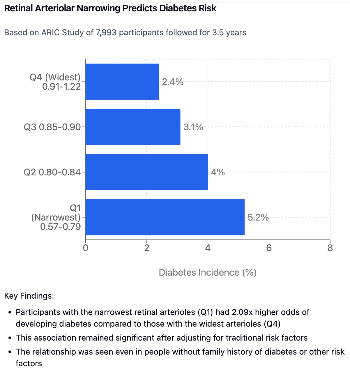
A landmark 2002 study published in JAMA 1 provided significant early evidence for fundus imaging’s potential in diabetes detection. The study, “Retinal Arteriolar Narrowing and Risk of Diabetes Mellitus in Middle-Aged Persons,” established the crucial link between retinal microvascular changes and diabetes risk. The researchers analyzed data from the Atherosclerosis Risk in Communities study, following 7,993 middle-aged participants without diabetes at baseline. By measuring the diameter of retinal arterioles and venules to calculate the arteriolar-to-venular ratio (AVR), they demonstrated that participants with narrower retinal arterioles faced a significantly increased risk of developing diabetes during the 3.5-year follow-up period. Those in the lowest quartile of the AVR ratio showed a 71% greater risk compared to those in the highest quartile, even after adjusting for traditional risk factors.
For modern clinical practice, these findings highlight fundus photography’s potential as a powerful tool for early and holistic health assessment. The ability to reveal early changes in retinal microvasculature before traditional diagnostic tests show abnormalities enables timely intervention and lifestyle modifications. Its non-invasive nature makes it particularly accessible and appealing to patients who might be hesitant about traditional medical procedures. The technology integrates seamlessly into broader health and wellness assessments, providing insights not only about ocular health but also systemic conditions. As a monitoring tool, sequential retinal images can track the progression of diabetes and treatment efficacy over time, enabling more targeted and effective care.
The connection between retinal health and diabetes is now well-established, and high-resolution fundus photography represents a remarkable opportunity to improve patient outcomes through early detection, enhanced monitoring, and a more comprehensive view of health. The integration of AI has democratized access to this technology, making sophisticated analysis available to a broad range of practitioners. This approach particularly excels in preventative and wellness care, offering valuable insights to both providers and patients while empowering individuals to take proactive control of their health journey.
2.5 Other Eye Pathologies
Glaucoma, another common ocular disease linked with various systemic factors, can also be identified by AI algorithms applied to fundus photos.2 These findings may have clinical impact because glaucoma is a frequent cause of blindness and can potentially be screened and treated earlier. Beyond this, some researchers have explored the link between thyroid disease and retinal fundus images and have found promising applications for diagnostic purposes, though further work is required.
Hypertensive retinopathy (HPR) manifests distinct patterns in fundus images that AI systems can now reliably detect. Research has shown strong correlations between the severity of HPR changes and systemic blood pressure levels. Advanced imaging analysis can quantify arteriolar narrowing, arteriovenous nicking, and other characteristic changes associated with HPR, providing valuable information about cardiovascular health risk.
Optic disc drusen, while often considered a benign finding, can sometimes be confused with more serious conditions like papilledema. AI-powered analysis helps differentiate these conditions by examining specific characteristics of the optic disc appearance. Studies have shown that machine learning algorithms can achieve high accuracy in distinguishing drusen from other optic disc abnormalities, helping guide appropriate clinical management.
Age-related macular degeneration (AMD) detection has been significantly enhanced by AI analysis of fundus images. Deep learning systems can now identify early signs of AMD, including subtle drusen formation and pigmentary changes, before they become clinically apparent. This early detection capability is crucial for implementing preventive measures and slowing disease progression.
Branch retinal vein occlusion (BRVO) and central retinal vein occlusion (CRVO) present distinct patterns in fundus images that AI systems can now identify with high reliability. Recent studies have demonstrated the ability of deep learning algorithms to detect these conditions early, potentially enabling faster intervention and better outcomes.
Recent advances in image analysis have also improved the detection of retinal artery occlusions. AI systems can now identify subtle changes in vessel caliber and perfusion patterns that might indicate impending occlusive events. This capability could help identify patients at risk for these sight-threatening conditions before they become clinically apparent.
The detection of optic neuritis through fundus imaging has also benefited from AI analysis. Machine learning algorithms can identify subtle changes in the optic disc appearance and peripapillary retinal nerve fiber layer that might indicate inflammatory or demyelinating processes. This capability has particular relevance for monitoring conditions like multiple sclerosis.
2.6 Retinal Imaging & Cardiovascular Health
The human eye increasingly appears to be a sophisticated mirror reflecting the overall health of the circulatory system. Within the retina, a delicate network of blood vessels—arterioles and venules—provides a unique, non-invasive opportunity to observe systemic vascular health. These microvessels, readily visible via non-mydriatic fundus photography, undergo subtle yet significant changes that are correlated with the increased risk of developing ischemic cardiovascular disease (ICVD). These changes, which include but are not limited to variations in arteriolar diameter, venular dilation, and the presence of microvascular damage, all indicate an underlying dysfunction within the body’s broader vascular system. In this section, we will explore the growing evidence linking retinal microvasculature and ICVD.
Traditional methods for assessing cardiovascular health, such as blood tests, blood pressure measurements, and questionnaires, provide essential but sometimes incomplete pictures of risk. These tests often require invasive procedures and/or complex interpretation and can be difficult to deploy at scale in community or primary care settings. Furthermore, risk assessment for CVD is still limited by the reliance on traditional risk factors, as many patients without these risk factors still develop heart disease. Retinal imaging, especially when combined with advanced image analysis and artificial intelligence (AI), offers a novel, non-invasive avenue for more direct and accessible assessment of a person’s vascular health and a tool that can be readily deployed in a wide range of clinical and community settings. One of the most compelling areas of research is the development of AI-driven approaches that are capable of predicting ICVD risk from retinal images, and these have shown remarkable performance in several large population studies.
One such study published in the Science Bulletin3, details how researchers from China utilized a vast dataset of over 390,000 retinal images to train a deep-learning algorithm for ICVD risk stratification. This study was based on non-mydriatic fundus images which makes them easy to collect in most clinical environments. The algorithm was designed to estimate a patient’s 10-year risk of ICVD events by learning to identify patterns in fundus images that may not be apparent to the naked eye, such as subtle changes in microvasculature. The model performed exceptionally well in both internal and external validation datasets, demonstrating robustness and generalizability across different groups of people. The model achieved an impressive adjusted R² of 0.876 on an internal data set and 0.638 on the external validation set which is the Beijing Research on Ageing and Vessel (BRAVE) data set. The adjusted R2 represents the proportion of variability that could be explained with this model. An R2 of 1 suggests that the model perfectly predicts outcomes with no variance, while 0 represents a model with no power to predict outcomes. These results show that AI-driven assessment of retinal imaging has high potential to estimate ICVD risk.
Furthermore, when using the trained algorithm to classify the risk of ICVD, the model showed a very high area under the receiver operating characteristic (AUC) curve for detecting patients with a 10-year ICVD risk of ≥5%. The AUC was 0.971 (95% CI: 0.967-0.975) in the internal validation dataset and 0.859 (95% CI: 0.822-0.895) in external validation. For the higher threshold of ICVD risk (≥ 7.5%), the AUC was 0.976 (95% CI: 0.973-0.980) for the internal validation dataset, and 0.876 (95% CI: 0.816-0.937) for external data. An AUC value close to 1 indicates perfect diagnostic accuracy. These AUC values demonstrate the high predictive power of this algorithm, which is consistent with other studies that have also seen a high predictive power of AI algorithms based on fundus images. The results indicate that this algorithm may be a feasible and accurate alternative to established methods for assessing risk of ICVD, which may lead to wide scale implementation of retinal imaging in routine check-ups. The findings also show that AI algorithms are able to learn the association of microvascular changes with ICVD, including venular dilatation and arteriolar narrowing. AI can extract subtle relationships from images which, while difficult to appreciate with the naked eye, can be predictive of health outcomes. These subtle changes are also consistent with other traditional risk factors, like blood pressure.
The study’s authors noted a few limitations. First, the data was collected cross-sectionally, and their outcomes were predicted from an estimation tool that used traditional risk factors, rather than actual longitudinal ICVD event data. To confirm the prediction ability, a follow-up study of the BRAVE data is planned. Second, smoking status was absent in the dataset. Despite the limitations, the findings still provide compelling evidence of AI’s potential in ICVD risk assessment using retinal images, given the simplicity of the approach and the high degree of predictive power.
2.7 Cerebral and Cognitive Health
The retina, during development, is an embryological extension of the brain, and as such shares an intimate physiological and anatomical relationship with it4. It’s an unusual tissue in that it can be observed non-invasively and allows an easy way to examine microvascular function. It is because of this that scientists are exploring the potential role of retinal imaging in understanding cerebrovascular and neurodegenerative diseases such as dementia. Retinal images provide a novel way to monitor cerebral health.
A growing body of research has established correlations between changes in the retinal vasculature and an increased risk of dementia. Studies have revealed that individuals with retinal microvascular abnormalities—including arteriolar narrowing, venular dilation, and the presence of retinopathy—have a higher likelihood of developing cognitive decline and dementia5. This link is rooted in the similarities between retinal and cerebral microvasculature. Both vascular systems share analogous structures and physiological functions, and changes in one may reflect similar pathological changes in the other. The implication of this relationship is important, because cerebrovascular disease is known to be a major contributor to dementia. Instead of solely relying on traditional cognitive tests, retinal imaging could be employed for population-wide screening, identifying high-risk patients earlier and allowing for earlier interventions.
In one innovative study6, researchers developed a novel algorithm utilizing fundus photographs to estimate the Cardiovascular Risk Factors, Aging, and Incidence of Dementia (CAIDE) dementia risk score. The CAIDE is a well-established tool that uses a multidimensional risk factors (age, sex, educational level, physical inactivity, systolic blood pressure, total cholesterol, and body mass index) to predict the 20-year risk of dementia. The study showed that the algorithm had a high adjusted R2 (0.80 in internal validation and 0.58 in external validation) for predicted CAIDE risk score compared with the actual score, suggesting the algorithm was able to extract the relevant data in the retinal photos. Furthermore, the external validation of the algorithm revealed a high area under the receiver operating characteristic curve (AUC) of 0.926 (95% CI: 0.913–0.939), indicating strong ability to discriminate individuals with high dementia risk. This predictive ability is very impressive, as CAIDE scores have also shown to be predictive in a large multiethnic population. This study moves beyond simple correlation and demonstrates that AI-driven analysis of retinal images can predict complex metrics associated with dementia risk, indicating a path for non-invasive early detection and risk stratification.
A similar study in China7 used fundus photographs from 271,864 participants across 19 regions, with external validation on 20,690 participants. The algorithm showed remarkable accuracy in identifying high dementia risk using the same CAIDE risk score, achieving an AUC of 0.944 in internal validation and 0.926 in external validation.
The algorithm demonstrated strong correlation between estimated and actual CAIDE scores, particularly in the internal validation group (R² = 0.80). Importantly, higher estimated risk scores were significantly associated with worse cognitive performance across multiple domains, confirming the clinical relevance of the predictions.
It’s important to point out that the study was cross-sectional rather than longitudinal, showed lower correlation in external validation (R² = 0.58), and was limited to a Chinese population. Additionally, the development dataset lacked complete lifestyle data that might have improved predictions.
Despite these limitations, this represents a significant advance in using retinal imaging for systemic health screening. The strong performance across different demographic groups and risk thresholds suggests this technology could help identify at-risk individuals for early intervention and clinical trial recruitment, though further validation with longitudinal outcomes data would be valuable.
Further supporting this connection between the retina and brain is work examining the impact of environmental factors on retinal structures. Researchers at the University College London analyzed the UK Biobank data set, and determined that exposure to ambient air pollution may be linked to changes in retinal layer thicknesses8. They found that increased exposure to fine particulate matter (PM2.5) and nitrogen oxides were correlated with a thicker retinal nerve fiber layer (RNFL) and a thinner ganglion cell-inner plexiform layer (GCIPL). Moreover, higher levels of PM2.5 absorbance were associated with a thinner RNFL, inner nuclear layer, and OPL+ONL. These findings not only suggest the impact of environmental toxins on retinal structure, but imply that these same toxins might also cause similar changes in other areas, including the brain.
Taken together, these investigations suggest that AI-based analysis of retinal images can potentially provide early, non-invasive indicators of brain health, providing a window into the pathological processes that may precede neurodegenerative conditions such as dementia.
2.8 Retinal Imaging & Anemia
Beyond its role as a window into vascular and neurological health, the retina also offers a unique opportunity for non-invasive assessment of hematological conditions such as anemia. Anemia, characterized by a deficiency in red blood cells or hemoglobin, affects an estimated 1.6 billion individuals worldwide and presents significant challenges in its diagnosis and management [1,2]. Due to the invasiveness and cost of diagnostic tests that require blood samples, the condition is often left undiagnosed, particularly in resource limited settings. However, the recent advances in AI, particularly when applied to retinal fundus photographs, offer a promising alternative for non-invasive detection and management of this important condition.
Researchers have demonstrated that AI algorithms can accurately quantify hemoglobin (Hb) levels and detect the presence of anemia using fundus photos alone. In a large-scale study published in Nature Biomedical Engineering9, a team of scientists used fundus images from the UK Biobank to develop deep learning models for the detection of anaemia using fundus photos, participant metadata or a combination of both. They found that a combined model of fundus images with metadata was most accurate, and the study used a validation set of 11,388 study participants. The results of the combined model showed a mean absolute error (MAE) of 0.63 g/dL (95% CI, 0.62–0.64) in predicted Hb concentration, an AUC of 0.88 (95% CI, 0.86-0.89) for anaemia detection, an area under the ROC curve of 0.88 (95% confidence interval (CI) 0.86-0.89) for detection of any anemia, and an area under the ROC curve of 0.95 (95% CI, 0.93-0.97) for moderate to severe anemia. The MAE of 0.63 g/dl was close to the accuracy of laboratory measurements of 0.14 g/dl (ref) and much more accurate than non-invasive point-of-care devices, whose accuracy is 1.1 to 1.2 g/dl. These results are striking because these outcomes are based entirely on non-invasive measurements. The fundus photos capture the subtle changes associated with low haemoglobin, including pallor of the retina and venous tortuosity. These findings not only highlight the capabilities of deep learning in processing complex image data but also show a clear path for a non-invasive method of diagnosing anaemia.
Moreover, the study also found that that the algorithm could detect anaemia in a group of 539 participants with self-reported diabetes, with comparable performance. The study had a slightly larger MAE of 0.73 g/dl (95% CI, 0.68-0.78 g/dl) and an AUC of 0.89 (95% CI, 0.85-0.93), as compared to all participants in the study. These results are particularly relevant because anemia is frequently associated with diabetes (up to 23% of patients with diabetes remain undiagnosed for anaemia) and is shown to increase morbidity and mortality in these populations. Given the potential for regular retinal screening of diabetic retinopathy, the capability of AI to also detect anemia from retinal photos can be of immense use and provide additional opportunities for healthcare screening.
2.9 Pupil Size and Intelligence
There is an intriguing correlation between baseline pupil size and cognitive ability, further supporting the eye’s role as a window into brain function. Studies conducted at the Georgia Institute of Technology10 have demonstrated that individuals with larger baseline pupil sizes tend to score higher on tests of fluid intelligence, attention control, and working memory capacity. This relationship proved robust enough that differences between high and low cognitive performers could be detected by the naked eye.
The physiological basis for this connection lies in the locus coeruleus, a nucleus in the upper brain stem that regulates pupil size and releases norepinephrine throughout the brain. This neurotransmitter plays a crucial role in perception, attention, learning, and memory. More importantly, it helps maintain organized brain activity across distant regions—a function essential for complex cognitive tasks.
The research team, using high-precision eye tracking technology, measured participants’ pupil sizes under controlled lighting conditions while they performed various cognitive assessments. Pupil diameters, which typically range from two to eight millimeters, showed consistent positive correlations with performance on fluid intelligence tests and attention control tasks. One particularly revealing test required participants to resist looking at a flickering stimulus and instead focus on identifying a briefly displayed letter—a task demanding sophisticated attention control.
This connection between pupil size and cognitive function appears to be age-dependent, with older participants generally showing more constricted pupils. However, when adjusted for age, the relationship between pupil size and cognitive ability remains significant. This finding suggests potential applications for non-invasive cognitive assessment through precise pupil measurement.
One hypothesis suggests that larger baseline pupil sizes indicate enhanced regulation by the locus coeruleus, potentially reflecting more efficient brain organization. This could explain the correlation with higher cognitive performance. Notably, dysfunction of the locus coeruleus has been implicated in conditions like Alzheimer’s disease and ADHD, suggesting potential diagnostic applications for careful pupil measurement in cognitive health assessment.
This research exemplifies how seemingly simple physiological measurements can provide windows into complex brain function. While more research is needed to fully understand these relationships, the findings reinforce the value of comprehensive ocular assessment in health evaluation. For practitioners using advanced imaging technology, awareness of these correlations can enhance their understanding of the eye’s role as a biomarker for overall health and cognitive function.
2.10 Predicting Age and Mortality Risk
While conventional wisdom might associate the retina solely with visual function, research is increasingly demonstrating that the eye also offers a window into the ageing process and a way to quantify mortality risk. The retina, composed of neural tissue and blood vessels, reflects both local changes that are influenced by age as well as the wider systemic effects of aging on the human body. Researchers have found that subtle age-related changes to the retina can be identified through fundus photography and quantified using AI, creating a novel biomarker of biological age and its connection with mortality risk.
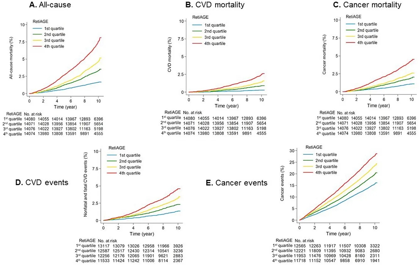
A team of researchers in Singapore developed an algorithm that can estimate a patient’s biological age, termed RetiAGE, based on deep learning from fundus images11. The algorithm was initially trained on fundus photographs from 40,480 Korean adults and then evaluated using 56,301 participants of the UK Biobank, which demonstrated its generalizability across diverse populations and ethnicities. They found that, using a cut off of being equal or greater than 65 years of age, the algorithm showed an AUC of 0.76, with an AUPRC of 0.399. More importantly, they then stratified participants by their RetiAGE and followed them for over 10 years and found that individuals in the fourth quartile of RetiAGE had a 67% increased risk of all-cause mortality, 142% increased risk of CVD-related mortality, and 60% increased risk of cancer related mortality compared to those in the lowest quartile. Critically, these associations were independent of chronological age and of a number of established ageing biomarkers including albumin, creatinine, glucose and C-reactive protein. This data suggests the algorithm is capturing some of the biological changes associated with aging that conventional biomarkers do not identify. In this study, the researchers also showed that the addition of RetiAGE increased the ability to predict mortality risk beyond the conventional risk factors.
Similarly, another study based on a 10 year longitudinal analysis of fundus images from the UK Biobank found that the retinal age gap (difference between predicted and chronological age) was associated with a 2% increase in all-cause mortality risk and 3% increased risk of non-CVD/non-cancer mortality12. While they did not find a significant association between retinal age gap and CVD or cancer-related mortalities, their findings underscore a role of retinal changes in broader ageing processes. Both the above studies have strong statistical significance with large populations and rigorous methodology, thus supporting the hypothesis that retinal fundus imaging could offer a non-invasive means of determining both biological age and risk of mortality.
While the biological mechanisms underlying the observed retinal changes associated with age and mortality remain the subject of future study, it is becoming increasingly evident that AI-driven analysis of retinal images can provide novel markers of both biological ageing and long-term health outcomes, demonstrating significant potential as a tool to assess mortality risk in a range of different settings.
2.11 Beyond The Main Focus
The body of evidence supporting the use of fundus photography for general health assessment continues to grow13, expanding beyond cardiovascular, neurological, and hematological conditions. AI is proving to be a versatile tool, and its capabilities in analyzing the complexity of retinal images are expanding our understanding of the retina and its link to a range of systemic diseases.
For example, research indicates potential applications in assessing liver function through retinal analysis. The retina’s unique vascular patterns may reflect subtle changes associated with hepatic conditions, as both organs share similar microvascular characteristics and regulatory mechanisms. Early studies suggest that specific retinal vascular patterns might correlate with liver enzyme levels and function, though more research is needed to validate these findings.
Emerging evidence also points to potential applications in immunological assessment. The retina’s immune-privileged status and its complex relationship with systemic immunity make it an interesting target for monitoring immune system function. Changes in retinal vasculature and tissue characteristics might provide early indicators of autoimmune conditions or immune system dysregulation.
Hormonal balance might be another area where retinal imaging could offer insights. The retina contains numerous hormone receptors, and preliminary research suggests that hormonal fluctuations may influence retinal vessel characteristics. This could potentially provide a non-invasive window into endocrine system function, though significant validation work remains to be done.
Researchers are also exploring connections between retinal patterns and gastrointestinal health. The gut-brain axis, which increasingly appears to influence various aspects of health, might manifest observable changes in retinal tissue. Some studies suggest that inflammatory bowel conditions could be reflected in retinal vascular patterns, though these findings are still preliminary.
The potential for retinal imaging to assess mitochondrial function represents another exciting frontier. Given the retina’s high metabolic demands and dense mitochondrial networks, changes in retinal tissue characteristics might reflect systemic mitochondrial health. This could have implications for understanding energy metabolism and aging-related conditions.
As analytical capabilities advance, researchers are investigating potential correlations between retinal features and microbiome health. While this connection might seem unlikely at first, emerging research suggests that gut microbiome composition could influence retinal health through systemic inflammatory pathways.
The field of chronobiology might also benefit from advanced retinal analysis. The retina’s role in circadian rhythm regulation suggests that detailed imaging could provide insights into circadian disruptions and their systemic effects. This could have implications for sleep medicine and metabolic health assessment.
These emerging areas of research highlight the continuing evolution of retinal imaging applications. As AI systems analyze larger datasets and identify new patterns, we’ll likely discover additional correlations between retinal features and various aspects of health. However, it’s important to maintain scientific rigor while exploring these new possibilities.
The future may reveal even more unexpected connections between retinal health and systemic conditions. As our understanding of the body’s interconnected systems deepens, the retina’s role as a window into overall health will likely expand. This underscores the importance of maintaining an open yet critical mindset when evaluating new applications for retinal imaging technology.
This ongoing research reinforces the value of incorporating retinal imaging into comprehensive wellness assessments. While some applications remain speculative, the growing body of evidence suggests that fundus photography will continue to reveal new insights into human health and disease processes.
These developments represent just the beginning of what may be possible with advanced retinal imaging. As technology continues to evolve and our understanding deepens, we can expect to discover additional applications that further enhance the value of this non-invasive assessment tool in wellness practice.